


In an innovative move, Harini Veeraraghavan, Ph.D., has been applying her tech skills to a crucial fight against cancer. At the Memorial Sloan Kettering Cancer Center (MSK), her lab is focused on creating advanced AI models aimed at making radiation therapy safer and more effective.Formerly, she contributed to projects like teaching robots to navigate indoors and designing computer programs for managing traffic.
The Challenge of Precision in Radiation Therapy
When it comes to radiotherapy, accuracy is everything. High-energy beams need to strike cancer cells with pinpoint accuracy; even a millimeter could mean impacting surrounding healthy tissue. This gets complicated as our organs can shift and change shape from day to day. “Breathing, food movement, or even liquids flowing can alter an organ’s shape,” Dr. Veeraraghavan states.
To address this complexity, she explains the need to create sophisticated computer models that account for these dynamic movements of our internal organs.
“We are in the process of building a virtual digital twin of a patient’s abdominal organs to strategize treatment better, ensuring radiation targets are accurately locked in,” Dr. Veeraraghavan notes.
Expanding AI’s Role Beyond Just the Abdomen
The research being done isn’t limited to just the abdomen but has implications for various cancers including head and neck, lung, and prostate cancers. “We are working with a team across multiple medical fields, ensuring that these cutting-edge AI technologies safely transition into everyday clinical use,” explains Joseph Deasy, Ph.D., Chair of Medical Physics at MSK.
Dr. Nancy Lee, MSK’s Chief of Head and Neck Radiation Oncology, highlights that these AI algorithms have drastically improved treatments for head and neck cancer patients by speeding up what used to be lengthy manual processes.
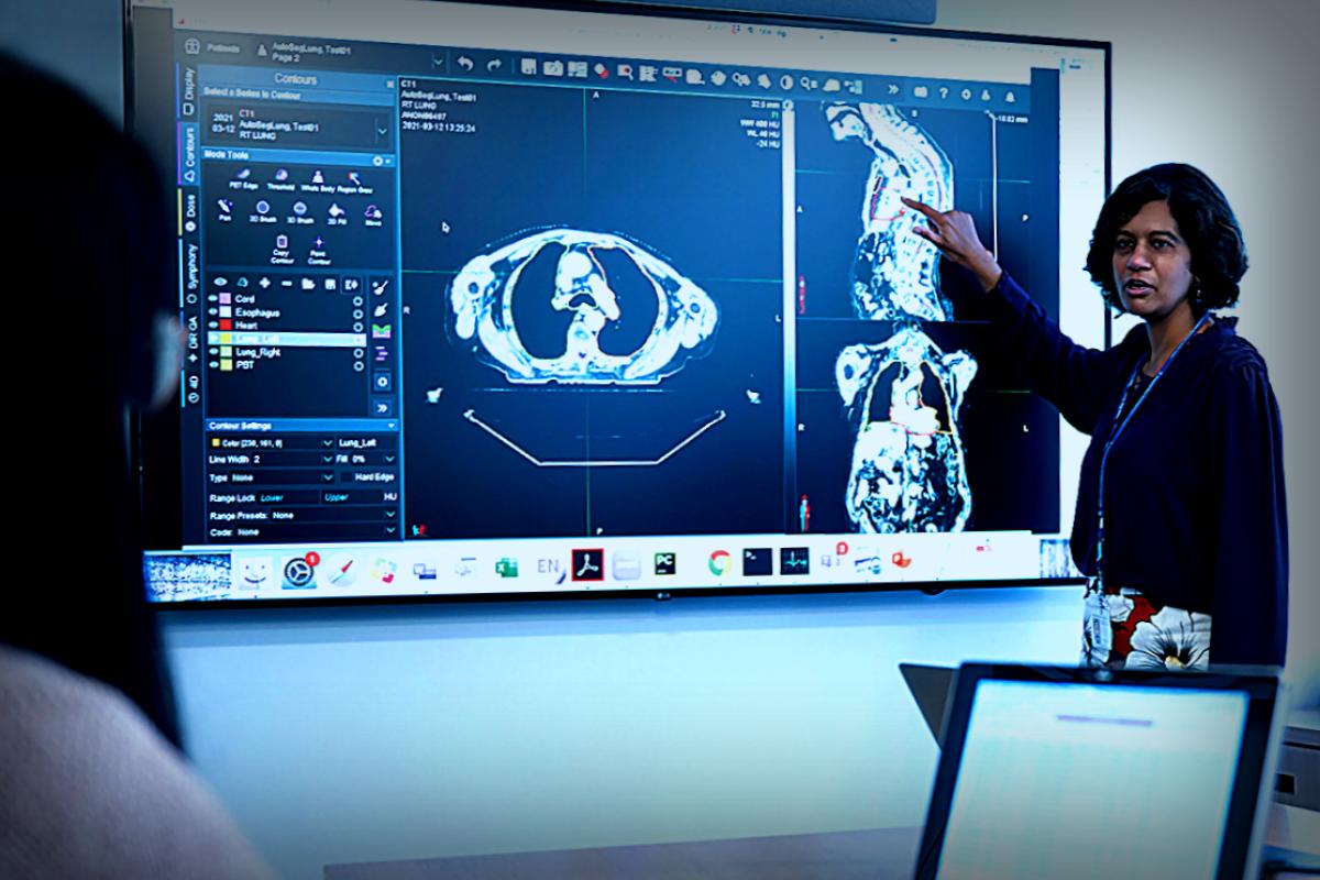
Traditionally, oncologists performed extensive manual contouring with CT or MRI scans to outline organs, which could take hours. Today, AI can generate these outlines automatically, allowing doctors to focus on making precise adjustments to ensure that cancerous tissues are accurately targeted while sparing healthy ones.
“It’s as if you had your best intern on the job,” Dr. Lee comments. “The algorithm can offer consistency and accuracy that even experienced oncologists may not replicate due to natural variation in human work.”
Using AI to Measure Tumors and Treatment Progress
But Dr. Veeraraghavan’s innovations don’t stop there. The technology is now assisting doctors in evaluating how tumors and lymph nodes are reacting to treatments more effectively. “These tools help us confirm whether to continue a therapy or switch to something better,” asserts radiologist Puneeth Iyengar, MD, Ph.D. “Previously, we had to wait months for results; now we can often predict outcomes far earlier.”
The conventional method for monitoring tumors is known as RECIST, which simply checks the measurement of a tumor’s diameter. The modern AI-equipped methods are capable of assessing a tumor’s volume in three dimensions, allowing for a much more comprehensive evaluation over time.
“It’s like measuring a body of water; simply taking width isn’t enough to know its overall dynamics,” Dr. Veeraraghavan adds.
Moreover, there are ongoing efforts with AI to track tumor hypoxia changes through standard PET scans, thus making assessing treatment resistance considerably more efficient. This helps doctors adapt radiation strategies based on concrete changes in the tumor behavior.
Initial insights gained from these measurements can lead to adjusting treatment plans promptly, which could even result in reduced radiation doses for effectively responding tumors, minimizing adverse effects for patients.
A Collaborative Effort Towards Future Innovations
Currently, these exciting AI-driven methodologies are being used in treating various cancers, including lung, brain, head, neck, and metastatic types, and efforts are underway to examine their applicability to prostate cancer.
Dr. Lee concludes that this work represents a collaborative effort, conveying a sense of camaraderie among a host of committed specialists. “It’s a team game that not only includes myself and Dr. Veeraraghavan but also valued contributions from many others at MSK,” she remarks.
This article originated from Memorial Sloan Kettering Cancer Center
Originally published on Medical Xpress.