


Recent research highlights the potential of perovskite detectors in enhancing high-energy scans, which are vital for spotting tumors and infections. These new detectors could replace the existing fragile and low-resolution systems, making diagnoses more precise while also minimizing radiation exposure, ultimately saving costs.
The advancements in nuclear medicine today allow us to assess biological activities within the body, like blood flow in tiny veins and arteries, unearthing abnormalities. These techniques are becoming more integral in brain imaging, helping doctors differentiate between types of dementia and navigate various treatment paths.
Professor Mercouri Kanatzidis from Northwestern University highlighted the remarkable versatility of perovskites, stating, “They revolutionized solar power technology, and now they’re on the brink of transforming nuclear medicine. Our findings confirm that perovskite detectors can provide high-quality images essential for effective patient care.” Learn more here.
Nuclear medicine encompasses treatments like radiotherapy, but in this scenario, the focus is on diagnostics. Single-photon emission computed tomography (SPECT) enables three-dimensional organ reconstructions by placing a gamma-ray source inside the body. The emitted rays—captured by detectors—help create complex images similar to X-rays, but delivered in a 3D format.
However, current detector technologies face challenges like high costs and issues with cracking (as seen with cadmium zinc telluride) or lackluster clarity (in sodium iodide). That’s where perovskites step in. Kanatzidis, a pioneer in perovskite solar technology, noted that the crystals produced by his team are fewer in imperfections, making them ideal for high-energy photon detection.
The landmark perovskite mineral starts with calcium titanium oxide, but now any crystal boasting the same structural traits qualifies. Kanatzidis discovered that CsPbBr perovskite displays perfect characteristics for advanced medical imaging.
“This work brilliantly showcases the tangible progress in deploying perovskite detectors beyond lab settings,” Kanatzidis remarked. “Our 2013 discovery showcased their capacity to detect X-rays and gamma rays, and now, we’re continuing to demonstrate their potential in key areas like nuclear medicine imaging.”
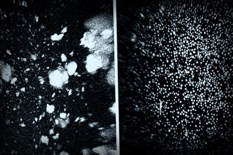
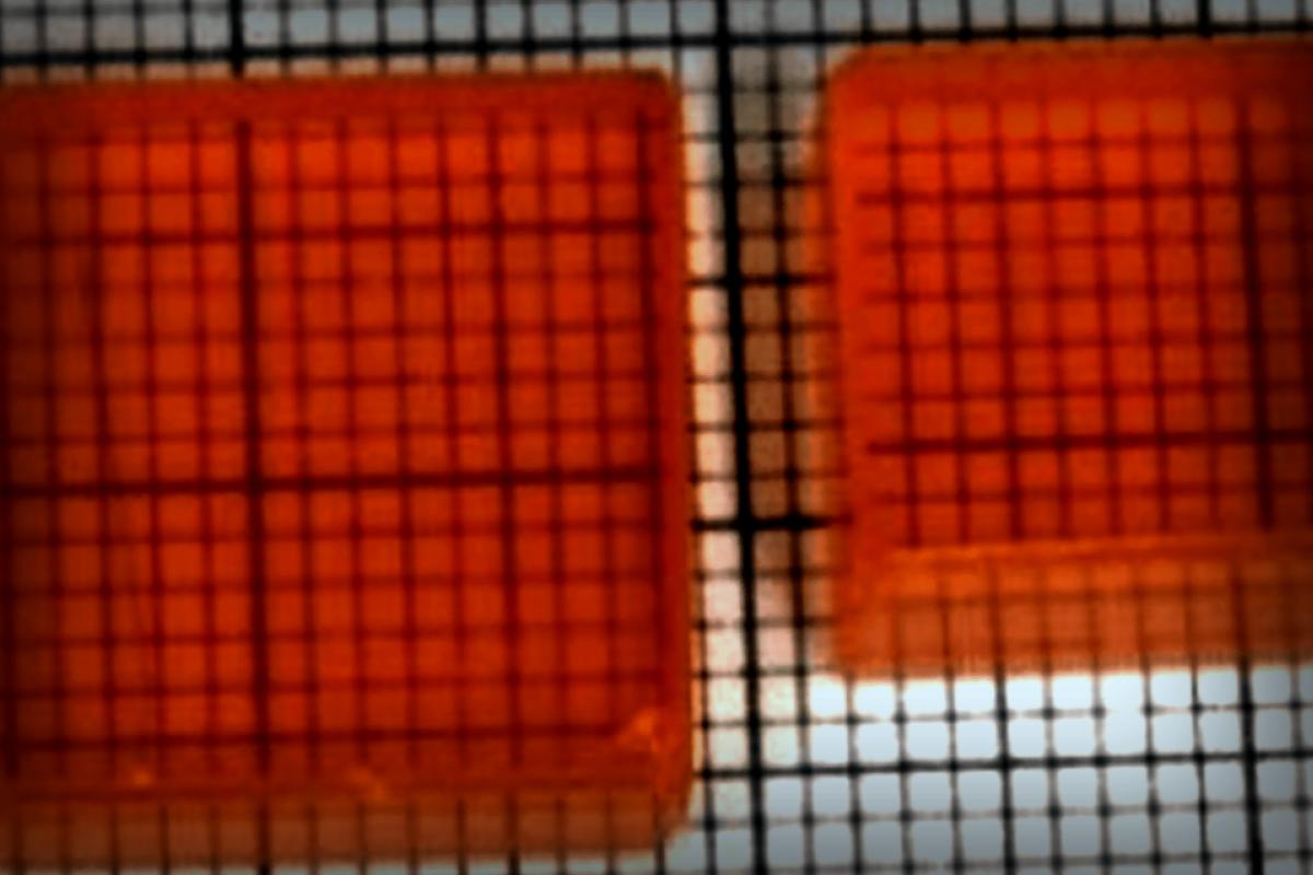
A collaboration between Kanatzidis and Professor Yihui He from Soochow University resulted in a pixelated gamma-ray sensor, capable of producing images with unprecedented resolution by capturing single gamma-ray photons. This capability enhances 3D organ imaging, especially differentiating gamma rays with varied energies, providing even more detailed and clearer images.
As a result, doctors can anticipate getting clearer images faster while minimizing necessary radiation doses when existing spots suffice. The cost-effectiveness and efficiency of making perovskites create buzz about their bright future, suggesting similar advancements for gamma-ray detectors should follow.
Now, Northwestern University has capitalized on this innovation by launching Actinia Inc., a company dedicated to advancing this groundbreaking technology. As professor He stated, “Proving that perovskites can enable single-photon gamma-ray imaging is a monumental step forward. It signals that these materials are ready to leap beyond research labs and into the clinical world, addressing significant health concerns. We’re on the cusp of scaling production and inventing new paths for medical imaging.”
Remember, if one day your life is saved by a scan that’s now financially accessible or has simply better resolution thanks to these advancements, you’ll owe it all to the merging of solar power research and medical science.
This study is accessible for everyone in Nature Communications.
Support Science Journalism. Stay connected!